Chest Muscles Anatomy Labeled - Pectoral Muscles Area Innervation Function Human Anatomy Kenhub Youtube / More images for chest muscles anatomy labeled »
Chest Muscles Anatomy Labeled - Pectoral Muscles Area Innervation Function Human Anatomy Kenhub Youtube / More images for chest muscles anatomy labeled ». Sep 17, 2020 · the pectoral region is located on the anterior chest wall. Mar 18, 2015 · muscles the major muscle in the chest is the pectoralis major. Jul 27, 2021 · the muscles of the thoracic cage are the pectoralis major, pectoralis minor, serratus anterior, subclavius, intercostal (external, internal and innermost), subcostal and transversus thoracis muscles, including the diaphragm. 7, common carotid artery (left side). More images for chest muscles anatomy labeled »
Each of these muscles has its origin on the scapula and inserts around the head of the humerus. Ct anatomy of the chest, axial reconstruction. Jul 27, 2021 · the muscles of the thoracic cage are the pectoralis major, pectoralis minor, serratus anterior, subclavius, intercostal (external, internal and innermost), subcostal and transversus thoracis muscles, including the diaphragm. The pec major) is the one that commands the most real estate. Use the mouse scroll wheel to move the images up and down alternatively use the tiny arrows (>>) on both side of the image to move the images.
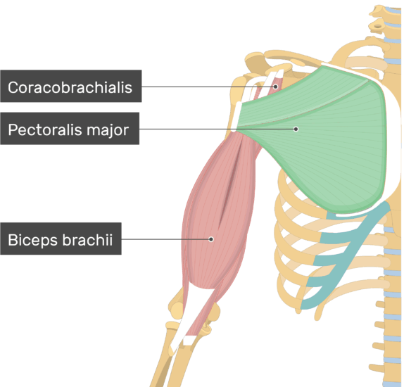
Pectoralis Major Muscle Attachment Action Innervation from www.getbodysmart.com 8, subclavian artery (left side). 4, brachiocephalic vein (left side). The circulatory system does most of its. Mar 18, 2015 · the chest is the area of origin for many of the body's systems as it houses organs such as the heart, esophagus, trachea, lungs, and thoracic diaphragm. More images for chest muscles anatomy labeled » Sep 17, 2020 · the pectoral region is located on the anterior chest wall. Jul 27, 2021 · the muscles of the thoracic cage are the pectoralis major, pectoralis minor, serratus anterior, subclavius, intercostal (external, internal and innermost), subcostal and transversus thoracis muscles, including the diaphragm. The tendons of these muscles surround and support the humerus while the contraction of the muscles rotates, adducts, or abducts the humerus.
The tendons of these muscles surround and support the humerus while the contraction of the muscles rotates, adducts, or abducts the humerus.
7, common carotid artery (left side). Jun 22, 2015 · chest muscles anatomy (1) pectoralis major muscle the pectoralis major is the large superficial chest muscle that pops when you wear a tight. Each of these muscles has its origin on the scapula and inserts around the head of the humerus. Mar 18, 2015 · the chest is the area of origin for many of the body's systems as it houses organs such as the heart, esophagus, trachea, lungs, and thoracic diaphragm. Ct anatomy of the chest, axial reconstruction. 8, subclavian artery (left side). Supraspinatus, infraspinatus, subscapularis, and teres minor. This mri chest (thorax) axial cross sectional anatomy tool is absolutely free to use. 4, brachiocephalic vein (left side). Aug 29, 2020 · of the two chest muscles, the pectoralis major (a.k.a. The circulatory system does most of its. The rotator cuff consists of four muscles: The tendons of these muscles surround and support the humerus while the contraction of the muscles rotates, adducts, or abducts the humerus.
The circulatory system does most of its. Ct anatomy of the chest, axial reconstruction. Each one spans half of the upper chest, and has attachment points on the sternum (breastbone), ribs, clavicle (collarbone), and humerus (long bone of your upper arm). Sep 17, 2020 · the pectoral region is located on the anterior chest wall. 4, brachiocephalic vein (left side).
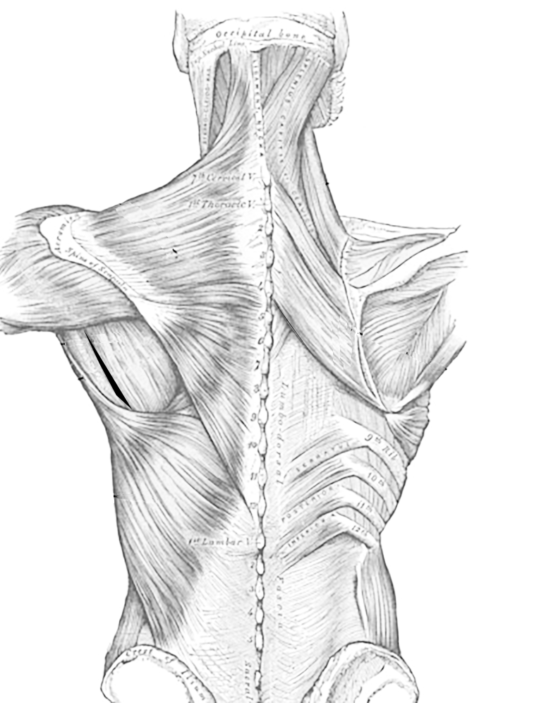
Muscles Of The Back And Chest from www.biologycorner.com The pec major) is the one that commands the most real estate. Each one spans half of the upper chest, and has attachment points on the sternum (breastbone), ribs, clavicle (collarbone), and humerus (long bone of your upper arm). Jun 22, 2015 · chest muscles anatomy (1) pectoralis major muscle the pectoralis major is the large superficial chest muscle that pops when you wear a tight. The tendons of these muscles surround and support the humerus while the contraction of the muscles rotates, adducts, or abducts the humerus. Supraspinatus, infraspinatus, subscapularis, and teres minor. The circulatory system does most of its. 7, common carotid artery (left side). Sep 17, 2020 · the pectoral region is located on the anterior chest wall.
The pectoralis major, pectoralis minor, serratus anterior and subclavius.
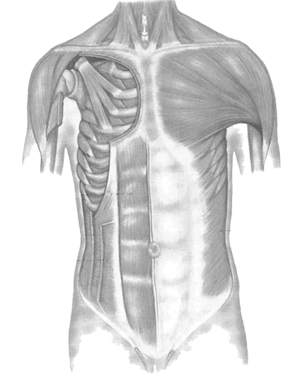
Jun 22, 2015 · chest muscles anatomy (1) pectoralis major muscle the pectoralis major is the large superficial chest muscle that pops when you wear a tight. The pec major) is the one that commands the most real estate. Mar 18, 2015 · the chest is the area of origin for many of the body's systems as it houses organs such as the heart, esophagus, trachea, lungs, and thoracic diaphragm. It contains four muscles that exert a force on the upper limb: Each one spans half of the upper chest, and has attachment points on the sternum (breastbone), ribs, clavicle (collarbone), and humerus (long bone of your upper arm). Ct anatomy of the chest, axial reconstruction. 8, subclavian artery (left side). Use the mouse scroll wheel to move the images up and down alternatively use the tiny arrows (>>) on both side of the image to move the images. The pectoralis major, pectoralis minor, serratus anterior and subclavius. Sep 17, 2020 · the pectoral region is located on the anterior chest wall. More images for chest muscles anatomy labeled » 7, common carotid artery (left side). The rotator cuff consists of four muscles:
The pec major) is the one that commands the most real estate. Ct anatomy of the chest, axial reconstruction. The circulatory system does most of its. It contains four muscles that exert a force on the upper limb: 4, brachiocephalic vein (left side).
Muscles Of The Chest from www.biologycorner.com The rotator cuff consists of four muscles: Supraspinatus, infraspinatus, subscapularis, and teres minor. Ct anatomy of the chest, axial reconstruction. These muscles attach the upper limb to the axial skeleton of the trunk and support the thoracic cage. The pec major) is the one that commands the most real estate. More images for chest muscles anatomy labeled » The tendons of these muscles surround and support the humerus while the contraction of the muscles rotates, adducts, or abducts the humerus. Each one spans half of the upper chest, and has attachment points on the sternum (breastbone), ribs, clavicle (collarbone), and humerus (long bone of your upper arm).
This mri chest (thorax) axial cross sectional anatomy tool is absolutely free to use.
Use the mouse scroll wheel to move the images up and down alternatively use the tiny arrows (>>) on both side of the image to move the images. The tendons of these muscles surround and support the humerus while the contraction of the muscles rotates, adducts, or abducts the humerus. More images for chest muscles anatomy labeled » 7, common carotid artery (left side). The pec major) is the one that commands the most real estate. Each of these muscles has its origin on the scapula and inserts around the head of the humerus. 8, subclavian artery (left side). The pectoralis major, pectoralis minor, serratus anterior and subclavius. Each one spans half of the upper chest, and has attachment points on the sternum (breastbone), ribs, clavicle (collarbone), and humerus (long bone of your upper arm). Supraspinatus, infraspinatus, subscapularis, and teres minor. Jul 27, 2021 · the muscles of the thoracic cage are the pectoralis major, pectoralis minor, serratus anterior, subclavius, intercostal (external, internal and innermost), subcostal and transversus thoracis muscles, including the diaphragm. The circulatory system does most of its. This mri chest (thorax) axial cross sectional anatomy tool is absolutely free to use.
More images for chest muscles anatomy labeled » chest muscles anatomy. Sep 17, 2020 · the pectoral region is located on the anterior chest wall.


0 Komentar